Course overview
The biological factors shaping the synaptic connectivity of neuronal circuits are complex and multifaceted, depending on cell types, functional activity, homeostasis, and more. Mapping brain wiring at the level of both local circuits and across brain-wide projections is a key aspect of understanding how nervous systems develop, learn, process information and generate behaviour. Recent advances in molecular biology, tissue processing, computational methods, and microscopy have enabled a revolution in understanding structural connectivity with cellular and synaptic resolution. Large-scale electron microscopy volumes provide nanometer-scale maps of anatomy and connectivity of whole invertebrate brains and millimetre-scale regions of vertebrate brains, while light-microscopic methods can highlight genetically defined connections and enable brain-wide reconstruction of neurons. Together, these complementary approaches yield powerful insight into the neuroanatomy and connectivity of the nervous system with single-cell resolution.
This course will provide students with a broad introduction to contemporary methods of studying neuronal connectivity with lectures from experts in the field. It also provides practical project-based instruction in experimental methods of circuit tracing and reconstruction with light microscopy (light sheet or 2-photon), as well as the computational analysis of rich electron microscopy connectomes in flies and mice. Students will consider the strengths and limitations of different techniques and how they can be used to address key problems in circuit neuroscience.
Course directors
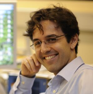
Gregory Jefferis
Course Director
MRC LMB and University of Cambridge, UK
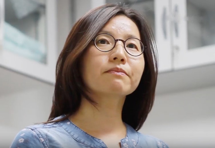
Jinny Kim
Course Director
Korea Institute of Science and Technology, Korea

Nicolas Renier
Course Director
Paris Brain Institute, France
Keynote Speakers
Jae-Byum Chang – KAIST, South Korea
Christel Genoud – University of Lausanne, Switzerland
Moritz Hemlstaedter – Max Plank Institute for Brain Research, Germany
Valentin Nägerl – University of Bordeaux, France
Alexandra Pacureanu – European Synchrotron Radiation Facility, France
Hiroki Ueda – Laboratory for Synthetic Biology, Japan
Claire Wyart – ICM Institute for Brain and Spinal Cord, France
Johannes Kohl – The Crick Institute, UK
Constantin Pape – University of Göttingen
Instructors / Teaching Assistants
Alba Vieites Prado – Universitad de Santiago de Compostela
Andrew Champion – Cambridge University, UK
Fabian Voigt – Harvard University, USA
In Cho – KAIST, Korean Advance Institute of Science and Technology, Korea
Jihyun Kim – KIST, Korean Institute of Science and Technology, Korea
Jordan Girard – université de Bordeaux
Katharina Eichler – Leipzig University, Germany
Leila Elabbady – University of Washington.
Martin Carbo-Tano – Paris Brain Institute, France
Philipp Schlegel – University of Cambridge, UK
Sahil Loomba – Max Planck Institute for Brain Research, Germany
Thomas Topilko – Gubra, Inc
Yagmur Yenner – Max Planck Institute for Brain Research, Germany
Yulia Dembitskaia – Université de Bordeaux
Course content
Light Microscopy and functional, molecular methods:
– Choice of labelling strategy: use of specific cre lines (for instance, the GENSAT project); finding specific markers for cell populations; using viral vectors and intersectional genetics (Dual or triple injections, transsynaptic tracing, Tango system, mGRASP, etc.).
– Tissue preparation for imaging: tissue clearing: choice of methods, considerations for the resolution needed and type of molecular labelling (Ueda, Renier); expansion microscopy methods: when to use them, and which iteration (Jae-Byum Chang)
– Imaging strategy: use of scanning microscopes: confocal or 2p, in intact samples or using serial sectioning; use of light sheet microscopy: commercial systems (eg. Miltenyi’s Blaze or Zeiss Z7), and custom systems (Mesospim).
– Analysis of imaging data: use of neuron mapping pipelines for whole brain data obtained from light sheet microscopy or from sections (eg. ClearMap, TrailMap, WholeBrain, etc). (Ueda, Renier); se of virtual reality-assisted tools for single neuron reconstructions from 3D datasets (eg. SyGlass, Vision4D…).
Electron microscopy synaptic connectomics:
At the end of this course, the students will be familiar with all of the steps that go into producing and analysing large scale, synaptic resolution EM connectomics datasets, summarised as below. Detailed analysis projects tailored by student interest will use public datasets and open source tools in which their directors and their colleagues are experts. These include the microns mouse cortical cubic millimetre dataset (https://www.microns-explorer.org/) and fly CNS datasets including the hemibrain, flywire.
Techniques
- EM imaging for connectomics (theory, image analysis)
- X-ray imaging for connectomics (theory, image analysis)
- EM connectomics data analysis (detailed hands on coverage for latest public whole brain fly and mouse cortex datasets; other organisms pending)
- In vivo calcium imaging
- In vivo 2-photon microscopy
- Light sheet microscopy
- Tissue Clearing
- Expansion Microscopy
- Brain mapping of cleared tissue (image analysis, commercial and academic softwares)
Projects
- Mapping of axonal projections with light sheet microscopy in the mouse brain
- Assembly of a Mesospim microscope for whole brain mapping with tissue clearing
- Brain mapping of cellular markers with HCR-fish, tissue clearing and light sheet microscopy
- In vivo recording of activity and connectivity in the Zebrafish larva
- Inference of information flow in Zebrafish larva using calcium imaging and Granger Causality
- Microscale connectomic analysis of mammalian connectomics data
- Morphological and connectivity analysis of the MICrONs mouse visual cortex dataset
- Student interest-led project(s) leveraging public mammalian connectomics data
- Whole brain circuit analysis using larval / adult Drosophila connectomes
- Student interest-led projects using public Drosophila connectomes
- Comparative connectomics: within species using multiple Drosophila datasets or across evolution using multiple public EM connectomes.
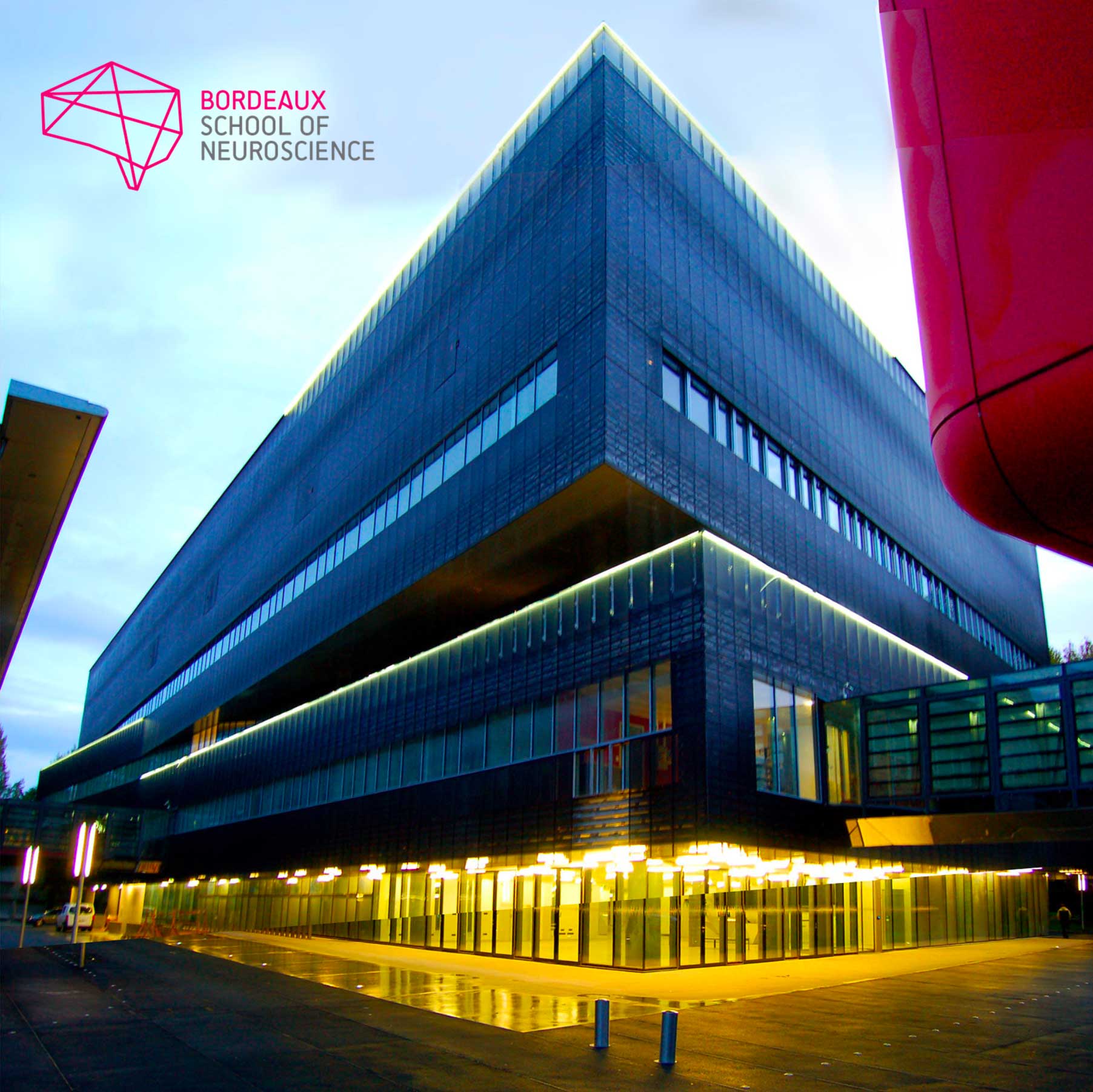
Bordeaux School of Neuroscience, France
The Bordeaux School of Neuroscience is part of Bordeaux Neurocampus, the Neuroscience Department of the University of Bordeaux. Christophe Mulle, its current director, founded it in 2015. Throughout the year, renowned scientists, promising young researchers and many students from any geographical horizon come to the School.
The school works on this principle: training in neuroscience research through experimental practice, within the framework of a real research laboratory.
Facilities
Their dedicated laboratory (500m2), available for about 20 trainees, is equipped with a wet lab, an in vitro and in vivo electrophysiology room, IT facilities, a standard cellular imaging room, an animal facility equipped for behavior studies and surgery and catering/meeting spaces. They also have access to high-level core facilities within the University of Bordeaux. They offer their services to international training teams who wish to organize courses in all fields of neuroscience thanks to a dedicated staff for the full logistics (travels, accommodation, on-site catering, social events) and administration and 2 scientific managers in support of the experimentation.
Registration
Fee : 3.950 € (includes tuition fee, accommodation and meals)
Applications closed 17 April 2023
The CAJAL programme offers 4 stipends per course (waived registration fee, not including travel expenses). Please apply through the course online application form. In order to identify candidates in real need of a stipend, any grant applicant is encouraged to first request funds from their lab, institution or government.
Kindly note that if you benefited from a Cajal stipend in the past, you are no longer eligible to receive this kind of funding. However other types of funding (such as partial travel grants from sponsors) might be made available after the participants selection pro- cess, depending on the course.


