Category Archives: On Site

Display this course using the On-Site template (blue)

Rescheduled: Advanced approaches to neuro evo-devo

This course has been rescheduled and will not be organised in 2022. Updates on the course status will be announced in the winter 2022.

Course overview

This course is a theoretical and practical training course on neurodevelopment and its evolution. It will provide an overview of the current concepts and knowledge on central nervous system development in several species, including invertebrates, mice, and humans, and their link to diseases. It will combine lectures and hands-on projects on convergent and divergent developmental processes across species at the molecular, cellular, circuit, and behavioural levels, including those relevant for human disease. It will include methods in genetics and molecular biology (e.g. genome editing), cellular neuroscience (e.g. transplantations), circuit neuroscience (e.g. live imaging) and -omics (e.g scRNA seq and bioinformatics).

This course will provide participants with a broad yet practical understanding of how the brain develops in different species, and how modern genetic approaches now allow cross-species comparisons to identify key developmental mechanisms. It is intended for PhD students and early-career postdocs.

Course directors
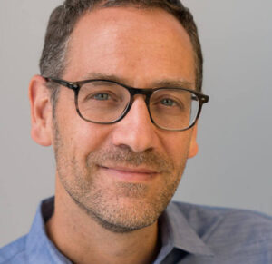
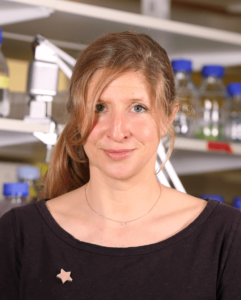
Denis Jabaudon

Denis Jabaudon

Course director

Faculty of Medicine,
University of Geneva, Switzerland
Paris Brain Institute, Institut du Cerveau (ICM), France
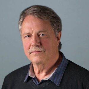
Claude Desplan

Claude Desplan

Co-director

Department of Biology, NYU New York, USA
Emilie Pacary

Emilie Pacary

Co-director

Neurocentre Magendie, University of Bordeaux, France

Keynote Speakers

Laure Bally-Cuif – Institut Pasteur, France
Sonia Garel – Ecole Normale Superieure (ENS), France
Simon Hippenmeyer – Institute of Science and Technology (IST), Austria
Oliver Hobert – Columbia University , USA
Guillermina Lopez Bendito – Instituto de Neurociencias – UMH-CSIC, Spain
Shubha Tole – Tata Institute of Fundamental Research, India

Instructors

Alexandre Baffet – Institut Curie, France
Nathan Benac – University of Bordeaux, France
Sara Bizzotto – Paris Brain Institute, France
Boyan Bonev – Helmholtz Zentrum München, Germany
Antoine de Chevigny – INMED, Univeristy of Marseille, France
Fernando García-Moreno, Achucarro Basque Center for Neuroscience, Bilbao, Spain
Juliette Godin – IGBMC Univeristy of Strasbourg, France
Isabel Holguera – New York University, USA
Nathalie Jurisch – Kavli Institute for Systems Neuroscience – NTNU, Norway
Karine Loulier – Institute for Neurosciences of Montpellier, France
Esther Klingler – UNIGE, Switzerland
Nikos Konstantinides – Institut Jacques Monod, France
Pierre Mortessagne – Neurocentre Magendie, Univeristy of Bordeaux, France
Homaira Nawabi – Grenoble Institute of Neuroscience, France
Stéphane Nédélec – Institut du Fer à Moulin, France
Sergi Roigg Puigros – UNIGE, Switzerland
Julie Stoufflet – Giga Liège, Belgium
Emre Yaksi – Kavli Institute for Systems Neuroscience – NTNU, Norway

Course content

Techniques

  • Crispr-CAS9 mediated genome editing to tag endogenous actin
  • Dissection and dissociation of mouse cortex
  • Fly husbandry
  • Human induced pluripotent stem cells culture
  • In utero/ex-utero electroporation
  • Analyses of cell position and protein expression using ImageJ
  • analyses of migration
  • ATAC-seq library preparation
  • Cell culture (dissociated hippocampal neurons, COS-7), cell transfection
  • Comparative neuroanatomy
  • Confocal microscopy and image analysis
  • Culture of organotypic brain slices
  • Drosophila brain dissection at L3 stage
  • Functional brain imaging in adult, juvenile and larval zebrafish and associated data analysis
  • Genetic manipulation of tTFs in neuroblasts using MARCM
  • Immunofluorescence
  • Image reconstructions and 3D analysis
  • Immunohistochemistry
  • Immunostaining
  • Intravitreal & in utero injections
  • Introduction to bioinformatics and scRNAseq analysis
  • Live birth-dating of neurons and functional imaging of their activity
  • Microtome / Vibratome sectioning
  • Retina, optic nerve dissection & explant culture
  • Spheroid generation and differentiation
  • Tissue freezing and cryostat slicing
  • Videomicroscopy,time lapse recording

See more techniques in the projects list.
Projects

  • Project 1 : “Live imaging of microtubule dynamics in mouse brain slices”
  • Project 2 : “Role of dopamine-glutamate interplay during synaptogenesis”
  • Project 3 : “Studying a developmental mosaic brain disorder in human cortical spheroids”
  • Project 4: “Histological and transcriptional analysis of isochronically labelled cortical neurons”.
  • Project 5: “Studying the morphological properties of developmentally- and adult-born dentate granule neurons”
  • Project 6: “RGC manipulation to modulate CNS regeneration”
  • Project 7: “Study of neuronal migration after prenatal alcohol exposure”
  • Project 8 : “Genome-wide profiling of the epigenome and the transcriptome of major cell types from the developing mouse cortex”
  • Project 9: “Emergence of intracortically-projecting neuron diversity”
  • Project 10 : “Studying the effect of temporal series in the proliferative capacity of Drosophila neural stem cells”
  • Project 11: “MAGIC Markers strategies to investigate neuron-astrocyte anatomical relationships during cortical development”
  • Project 12: “ Differentiation of spinal organoids and motor neuron subtypes from human induced pluripotent stem cells”
  • Project 13: “Inferring ligand-receptor interactions between GABAergic and Glutamatergic cells during Somatosensory Cortex Development”
  • Project 14: “Imaging actin cytoskeleton dynamics during radial migration of projection neurons in the mouse developing cortex”.
  • Project 15: “Neurogenesis in the Drosophila optic lobe by temporal patterning”
  • Project 16: “Fluid dynamics in the brain of zebrafish and medaka larvae”
  • Project 17: “Imaging function, connectivity, and development of brain circuits in zebrafish”
  • Project 18: “Comparative neurodevelopment of mouse and chick brains”
Bordeaux School of Neuroscience, France

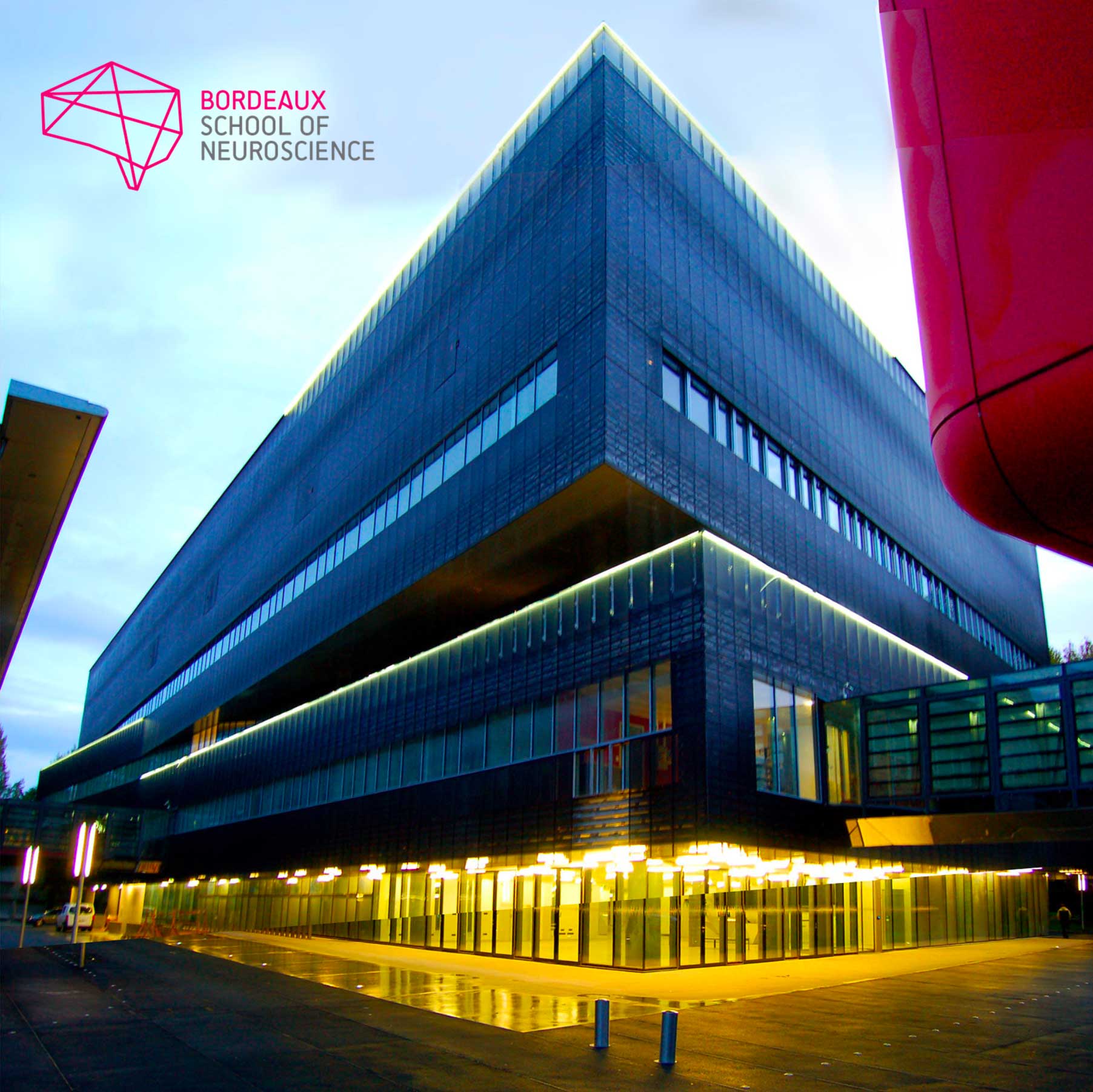
The Bordeaux School of Neuroscience is part of Bordeaux Neurocampus, the Neuroscience Department of the University of Bordeaux. Christophe Mulle, its current director, founded it in 2015. Throughout the year, renowned scientists, promising young researchers and many students from any geographical horizon come to the School.
The school works on this principle: training in neuroscience research through experimental practice, within the framework of a real research laboratory.

Facilities
Their dedicated laboratory (500m2), available for about 20 trainees, is equipped with a wet lab, an in vitro and in vivo electrophysiology room, IT facilities, a standard cellular imaging room, an animal facility equipped for behavior studies and surgery and catering/meeting spaces. They also have access to high-level core facilities within the University of Bordeaux. They offer their services to international training teams who wish to organize courses in all fields of neuroscience thanks to a dedicated staff for the full logistics (travels, accommodation, on-site catering, social events) and administration and 2 scientific managers in support of the experimentation.

Registration

Fee : 3.500 € (includes tuition fee, accommodation and meals)

The CAJAL programme offers 4 stipends per course (waived registration fee, not including travel expenses). Please apply through the course online application form. In order to identify candidates in real need of a stipend, any grant applicant is encouraged to first request funds from their lab, institution or government.

Kindly note that if you benefited from a Cajal stipend in the past, you are no longer eligible to receive this kind of funding. However other types of funding (such as partial travel grants from sponsors) might be made available after the participants selection pro- cess, depending on the course.

Brain Organoids

Course overview

Recent advancement in the stem cell field has led to the development of novel 3D cell culture models called organoids that mimic cell type diversity and architecture during organogenesis. Brain organoids, derived from human embryonic stem cells or induced pluripotent stem cells, capture key features of the developing human brain, including stem cell pool expansion, neurogenesis, gliogenesis, synaptogenesis and cytoarchitecture formation with cellular diversity and complexity. Organoids can also be derived from patient tumor samples, such as glioblastoma, for modelling brain tumors. In less than a decade, brain organoids have already been shown to be an extremely valuable tool to understand the human brain, and novel insights have been gained in deciphering evolution, human-specific features related to the brain development and neurological diseases resulted from pathogen infection, environmental insult, or genetic mutations.

In this course, we will take a multi-disciplinary approach to show what we could learn from brain organoid technology and what the future holds. The keynote speakers are all leaders in the field and will showcase the most up-to-date knowledge of brain organoids.

Course directors

Guo-Li Ming

Course director

University of Pennsylvania, USA

Hongjun Song

Co-director

University of Pennsylvania, USA

Marisa Karow

Co-director

Friedrich-Alexander University of Erlangen-Nürnberg, Germany

Keynote Speakers

Alex Baffet – Institute Curie, Paris, France
Gray Camp – Roche Institute, Basel, Switzerland
Silvia Cappello – MPI Psychaitry, Munich, Germany
Mike Karl – CRTD, Dresden, Germany
Agnete Kirkeby – reNEW, Copenhagen, Denmark
Jürgen Knoblich – IMBA, Vienna, Austria
Matthias Lütolf – Roche Institute, Basel, Switzerland
Abed Mansour – The Hebrew University of Jerusalem, Israel
Sergiu Pasca – Wu Tsai Neurosciences Institute, Stanford, US
Lorenz Studer – Memorial Sloan-Kettering Cancer Center, US
Barbara Treutlein – ETH, Zurich, Switzerland

Instructors

Giovanna Brancati – BSSE, ETH Zürich, Basel, Switzerland
Clarisse Brunet – Institut Curie, Paris, France
Maren Büttner – DZNE, Bonn, Germany
Francesco Di Matteo – Max Planck Institute for Psychiatry, Munich, Germany
Sarah Frank – FAU Erlangen-Nürnberg, Institute of Biochemistry, Erlangen, Germany
Federica Furlanetto – FAU Erlangen-Nürnberg, Institute of Biochemistry, Erlangen, Germany
Yan Hong – Perelman School of Medicine at the University of Pennsylvania, Philadelphia, USA
Richard O’Laughlin – Perelman School of Medicine at the University of Pennsylvania, Philadelphia, USA
Laura Pellegrini – MRC Laboratory of Molecular Biology, Cambridge, UK
Fides Zenk – BSSE, ETH Zürich, Basel, Switzerland
Ting Zhao – Perelman School of Medicine at the University of Pennsylvania, Philadelphia, USA

Course content

Topics & Techniques

The following techniques will be taught at the course:

• Generation of organoids and assembloids
• Organoids characterization
• CSF sampling from choroid plexus organoids
• Viral infection (transduction)
• Live-cell imaging (spinning-disk confocal microscope)
• Setting up of a microfluidic device
• Evaluation of the performance of microfluidic devices
• Extracellular multielectrode array (MEA) recordings
• FACS sorting
• Single cell RNA sequencing
• Generation of retina organoids
• Single cell analysis
• SCANPY framework
• Confocal microscopy
• Surgical procedure of organoid transplantation
• Animal perfusion
• Immunofluorescence
• Image analysis

brain organoids

Projects

Project 1: Generation and characterization of brain organoid of different regional identities and assembloids derived
Project 2: Microfluidic methods for patterning brain organoids
Project 3: Different approaches to perform Electrophysiological recordings in mature human cerebral organoids
Project 4: Using single cell RNA sequencing to decipher cellular heterogeneity of cerebral organoids
Project 5: Transplantation of hPSC-derived brain organoids into mouse brain
Project 6: A sneak peek into retinal organoids
Project 7: Generation and characterization of specialized organoids: cerebral and choroid plexus organoids
Project 8: Revealing neuronal activity in brain organoids using microelectrode array (MEA)
Project 9: Single cell analysis in brain organoids
Project 10: Investigate the Neurogenesis of Transplanted Forebrain Organoids In Vivo with Immunostaining
Project 11: To Fate or Not To Fate: live-cell imaging of neural progenitor cells to study cell fate decisions in the human developing neocortex

Get more information on the projects in the projects list file below.

Bordeaux School of Neuroscience, France

The Bordeaux School of Neuroscience is part of Bordeaux Neurocampus, the Neuroscience Department of the University of Bordeaux. Christophe Mulle, its current director, founded it in 2015. Throughout the year, renowned scientists, promising young researchers and many students from any geographical horizon come to the School.
The school works on this principle: training in neuroscience research through experimental practice, within the framework of a real research laboratory.

Facilities
Their dedicated laboratory (500m2), available for about 20 trainees, is equipped with a wet lab, an in vitro and in vivo electrophysiology room, IT facilities, a standard cellular imaging room, an animal facility equipped for behavior studies and surgery and catering/meeting spaces. They also have access to high-level core facilities within the University of Bordeaux. They offer their services to international training teams who wish to organize courses in all fields of neuroscience thanks to a dedicated staff for the full logistics (travels, accommodation, on-site catering, social events) and administration and 2 scientific managers in support of the experimentation.

Registration

Fee : 3.500 € (includes tuition fee, accommodation and meals)

Applications closed on 30 May 2022

The CAJAL programme offers 4 stipends per course (waived registration fee, not including travel expenses). Please apply through the course online application form. In order to identify candidates in real need of a stipend, any grant applicant is encouraged to first request funds from their lab, institution or government.

Kindly note that if you benefited from a Cajal stipend in the past, you are no longer eligible to receive this kind of funding. However other types of funding (such as partial travel grants from sponsors) might be made available after the participants selection pro- cess, depending on the course.
Course sponsors

The Tianqiao and Chrissy Chen Institute is a science foundation dedicated to advancing our understanding of the full complexity of the brain and mind. The foundation’s mission is implemented through partnerships with world-leading universities and major scientific societies.

At Scientifica, we employ experience, collaboration, and superior design to empower you to discover the brain’s secrets and overcome neurological diseases. Our equipment is optimised for electrophysiology, multiphoton imaging and optogenetics studies.

Our highly qualified team provides first-class service and support, and our resources centre is packed with invaluable and educational content. Get in touch to see how we can help you achieve your research goals.

Optogenetics, chemogenetics and biosensors for cellular and circuit neuroscience

Course overview

Spatio-temporally precise manipulation and read-out of brain circuit function has been one of the longest-standing challenges in neuroscience. The recent explosion in the field of genetically encoded tools to control and measure neuronal activity has greatly facilitated investigation of brain function, ranging from single synapses to large-scale circuits. Both control and readout of neuronal activity can now be achieved over orders of magnitude in space and time, ranging from micrometers to entire brain regions and from milliseconds to days.

This course will provide participants with the opportunity to gain hands-on experience using the latest genetically encoded tools and state-of-the-art equipment for brain circuit investigation. A particular focus will lie on multiplexed manipulations and read-out of brain circuits. Participants will be familiarized with the biophysical principles behind the sensors and actuators, and given training complementary to their background in the technical aspects of experimental approaches.

Hands-on experiments will employ optogenetic and chemogenetic actuators, including excitatory and inhibitory ion channels, pumps, enzymes and G-protein coupled receptors. These actuators will be complemented by genetically encoded indicators of neural activity, including calcium and voltage indicators as well as indicators for neurotransmitters and neuromodulators such as glutamate, dopamine and norepinephrine.

The course will cover a wide range of experimental systems with an emphasis on functional brain circuits in vivo. Finally, participants will be guided through data analysis and conceptual interpretations of their experiments.

Course partner

Course directors

Ofer Yizhar

Course Director

Weizmann Institute of Science

Israel

Michael Lin

Co-director

Stanford University

USA

Simon Wiegert

Co-Director

Center for Molecular Neurobiology Hamburg (ZMNH)

Germany

Anna Beyeler

Co-director

University of Bordeaux

France

Keynote speakers

Adam Cohen – Harvard University, USA
Stephane Dieudonne – University of Marseille, France
Valentina Emiliani – Institut de la vision, France
Stefan Herlitze – University of Bochum, Germany
Na Ji – University of Berkeley, USA
Tom Kash – University of North Carolina, USA
Tatiana Korotkova – MPI Köln, Germany
Adam Packer – University of Oxford, UK
Tommaso Patriarchi – ETH Zurich, Switzerland
Jonas Wietek – Weizmann Institute of science, Israel
Yaniv Ziv – Weizmann Institute of science, Israel

Instructors

Yoann Atlas – Institut de la vision, France
Imane Bendifallah – Institut de la vision, France
Miklos Boldogkoi – Institute of Molecular and Clinical Ophthalmology Basel IOB, Switzerland
Martina De Gennaro – Institute of Molecular and Clinical Ophthalmology Basel IOB, Switzerland
Alexander Dieter – Center for Molecular Neurobiology Hamburg – ZMNH, Germany
Andrey Formozov – Center for Molecular Neurobiology Hamburg ZMNH, Germany
Nitzan Geva – Weizmann Institute of science, Israel
Christianne Grimm – Institut de la vision, France
J. Quinn Lee – McGill University, Canada
Maxime Maheu – University Medical Center Hamburg-Eppendorf, Germany
Mathias Mahn – Friedrich Miescher Institute of Basel, Switzerland
Vasyl Mykytiuk – Max Plank Institute, Germany
Praneeth Namburi – Massachusetts Institute of Technology, USA
Mauro Pulin – Center for Molecular Neurobiology Hamburg – ZMNH, Germany
Inbar Saraf-Sinik – Weizmann Institute of science, Israel
Robson Scheffer Teixeira – University of Cologne, Germany
Guilherme Silva – University of Harvard, USA
Dimitrii Tanese – Institut de la vision, France

Course content

Topics & Techniques

The following techniques will be covered during the course:

  • implantation of optical fibers, stereotactic injection of viruses in mice.
  • optogenetic stimulation (BIPOLES) and monitoring of pupil size and mouse behavior.
  • imaging of calcium responses, building of a reward delivery system and a head-fixation system for mice either immobilized in a tube or running on a treadmill.
  • in vitro exploration of FLARE technique
  • 2-photon holographic illumination to achieve single-cell resolved optogenetic activation of presynaptic cells and patch-clamp recording of the post-synaptic neuron
  • concurrent photostimulation and calcium imaging of the presynaptic cells, and voltage imaging on the post-synaptic cell
  • use of miniature head-mounted microscopes (Inscopix).
  • perform population level data analysis on data that was collected across multiple days
  • behavioral assays including the elevated plus maze, the open field test, consumption of water, sucrose, quinine and food, as well as mild foot shocks
  • head-bar implantation, stereotaxic intracerebral virus injection, and craniotomy preparation for long-term recordings
  • head-fixed electrophysiology setup building
  • silicon-probe recordings, in head-fixed mice during behavior, using multi-shank, high-density silicon probes (128-512 channels) & Physiological monitoring
  • opto-tagging and optogenetic manipulations of specific cell-types.
  • open-source hardware and software for the acquisition and processing of the data, including OpenEphys, Bonsai, PulsePal, Cyclops, Arduino, Linux, KiloSort, and MountainSort
  • construct microdrives, mount the silicon probes onto them, make optical fibers of custom length

Projects

  • Project 1: “Optogenetic control of neuromodulation ”
  • Project 2: “Optical tool exploration in culture, in slice, and in vivo”
  • Project 3: “Optical tool exploration in culture, in slice, and in vivo”
  • Project 4: “Probing neuronal excitability with Arch-derived voltage sensors ”
  • Project 5: “Longitudinal calcium imaging in freely behaving mice using Inscopix system”
  • Project 6: “In vivo imaging of divergent neural populations using dual-color fiber photometry”
  • Project 7: “All optical characterization of eOPN3 mediated terminal inhibition in vivo”
  • Project 8: “Large-scale electrophysiology and optogenetics during head-fixed behavior”
  • Project 9: “In vivo calcium imaging with open-source Miniscopes”
  • Project 10: “Combining in vivo electrophysiology and optogenetics in freely moving mice”
  • Project 11: “All optical interrogation of dopamine circuits in freely moving mice using multiplexfiber photometry and biosensors”
  • Project 12: “All-optical manipulation and read-out of synaptic transmission”

For more information on projects and techniques which will be taught at the course, download the projects list.

Bordeaux School of Neuroscience, France

The Bordeaux School of Neuroscience is part of Bordeaux Neurocampus, the Neuroscience Department of the University of Bordeaux. Christophe Mulle, its current director, founded it in 2015. Throughout the year, renowned scientists, promising young researchers and many students from any geographical horizon come to the School.
The school works on this principle: training in neuroscience research through experimental practice, within the framework of a real research laboratory.

Facilities
Their dedicated laboratory (500m2), available for about 20 trainees, is equipped with a wet lab, an in vitro and in vivo electrophysiology room, IT facilities, a standard cellular imaging room, an animal facility equipped for behavior studies and surgery and catering/meeting spaces. They also have access to high-level core facilities within the University of Bordeaux. They offer their services to international training teams who wish to organize courses in all fields of neuroscience thanks to a dedicated staff for the full logistics (travels, accommodation, on-site catering, social events) and administration and 2 scientific managers in support of the experimentation.

Registration

Fee : 3.500 € (includes tuition fee, accommodation and meals)

Application closed on 7 December 2020

The CAJAL programme offers 4 stipends per course (waived registration fee, not including travel expenses). Please apply through the course online application form. In order to identify candidates in real need of a stipend, any grant applicant is encouraged to first request funds from their lab, institution or government.

Kindly note that if you benefited from a Cajal stipend in the past, you are no longer eligible to receive this kind of funding. However other types of funding (such as partial travel grants from sponsors) might be made available after the participants selection pro- cess, depending on the course.
COVID-19 update: In case the Optogenetics, chemogenetics and biosensors for cellular and circuit neuroscience course is postponed due to the pandemic, all applicants will have the choice to maintain their application or cancel it. Applicants who were already selected to attend will not have to reapply and will automatically be enrolled in the rescheduled course.
In addition, the Cajal Programme will not, as far as possible, request the registration fee from selected applicants until the course has been secured and confirmed. Nevertheless, should the course be cancelled before the course dates and the registration fees already collected, participants will be reimbursed.

Course sponsors

Neuroepigenetics: writing, reading and erasing the epigenome

Course overview

This course is a theoretical and practical training on the recently emerged field of neuroepigenetics. It will provide an overview of the current concepts and knowledge on the nature and functions of the epigenome in the nervous system, its modes of regulation and its link to brain health and disease. It will combine lectures and hands-on projects to learn about state-of-the-art approaches and methodologies to study how the epigenome is established and modulated by behaviour in rodents and invertebrates, what machinery is involved and what is its causal relationship to functions. It will include methods in behaviour, epigenetics, (epi)genome editing, molecular and cell biology, -omics and bioinformatics.

This course wishes to foster the development of neuroepigenetics as a key discipline in the neurosciences, and train and educate young researchers expected to contribute to the field in the near future. It is intended for PhD students and early career postdocs who wish to acquire good bases and broad knowledge of the field.

Course partner

Course directors

Karine Merienne

Course Director

CNRS – LNCA, University of Strasbourg, France

Angel Barco

Co-Director

Neurosciences Institute (UMH-CSIC), Spain

André Fischer

Co-Director

German Center for Neurodegenerative diseases (DZNE), Germany

Institute of Informatics, University of Warsaw, Poland

Keynote Speakers

Elisabeth Binder – Max Planck Institute of Psychiatry, Munich, Germany
Anne-Laurence Boutillier – LNCA, Strasbourg, France
Goncalo Castelo Branco – Karolinska Institute, Stockholm, Sweden
Johannes Gräff – EPFL, Lausanne, Switzerland
Elizabeth A Heller – Penn Epigenetics Institute, Philadelphia, USA
Denes Hnisz – Max Planck Institute for Molecular Genetics, Berlin, Germany
Aleksandra Pekowska – Dioscuri Center of Chromatin Biology and Epigenomics, Poland

Instructors

Rafael Alcala Vida – Instituto de Neurociencias UMH-CSIC, Spain
Mykhailo Batiuk – EPFL, Lausanne, Switzerland
Davide Coda – EPFL, Lausanne, Switzerland
Charles Decraene – LNCA, Strasbourg, France
Beatriz del Blanco Pablos – Instituto de Neurociencias UMH-CSIC, Spain
Aleksander Jankowski – Institute of informatics, Warsaw, Poland
Lalit Kaurani – DZNE, Göttingen, Germany
Marta Kullis – IDIBABS, Barcelona, Spain
Stéphanie Legras – IGBMC, Strasbourg, France
Jose Lopez Atalaya – Instituto de Neurociencias UMH-CSIC, Spain
Pierre-Eric Lutz – INCI, Strasbourg, France
Magdalena Machnicka – Institute of informatics, Warsaw, Poland
Marta Alaiz Noya – Instituto de Neurociencias UMH-CSIC, Spain
Isabel Paiva – LNCA, Strasbourg, France
Jose Sanchez-Mut – Instituto de Neurociencias UMH-CSIC, Spain
Sophie Schroeder – DZNE, Göttingen, Germany
Tanya Vavouri – Pujol Research Institute, Badalona, Spain

Course content

Topics & Techniques

State-of-the-art approaches to produce and analyze cell-type-specific epigenomic data in the brain in physiological and disease contexts will be taught, as well as approaches to manipulate the epigenome in brain cells.

  • ChIP seq
  • CUT & Tag seq
  • ATAC seq
  • Single nuclei DNA methylome (snmC-seq2)
  • Single nuclei RNA seq
  • Sample preparation for simultaneous RNAseq, WGBSseq and/or singe-nuclei ATAC-RNAseq (multiome)
  • Design, production and manipulation of epi-editing tools using viral constructs and CRISPR system, and analysis of transcriptional changes at targeted loci
  • Mouse brain subregion dissection, tissue sectioning
  • Fluorescently activated nuclear sorting (FANS) of neuronal and non-neuronal cell populations from mouse brain tissue
  • Isolation of glial cells (astrocytes) from mouse brain tissue using magnetic beads (MACS)
  • RNAscope
  • Confocal microscopy
  • Primary cultures of hippocampal neurons.

Projects

Project 1: Cell-type-specific epigenetic profiling using Fluorescence-Activated Nuclear Sorting (FANS) from brain tissue

Project 2: Astrocyte epigenome of mouse hippocampus

Project 3: Transcriptomic profiling of adult mouse brain tissue using 10X Genomics single-nuclei RNA-sequencing

Project 4: Cell type-specific detection of lncRNAs in the mouse brain using RNAscope and immunofluorescence

Project 5: Investigation of single cell DNA methylome landscape of cortical brain cells

Project 6: Cell-type and brain-region specific epigenetic manipulation in Alzheimer’s disease models

Project 7: CRISPR-dCas9 systems for targeted epigenetic repression

Project 8: Epigenome editing technology based on the CRISPR/dCas9 system.

Computational projects: State-of-the-art approaches for bioinformatic analysis and interpretation of the next generation sequencing (NGS) data obtained in the epigenomic experiments covered in the first block.

Bordeaux School of Neuroscience, France

The Bordeaux School of Neuroscience is part of Bordeaux Neurocampus, the Neuroscience Department of the University of Bordeaux. Christophe Mulle, its current director, founded it in 2015. Throughout the year, renowned scientists, promising young researchers and many students from any geographical horizon come to the School.
The school works on this principle: training in neuroscience research through experimental practice, within the framework of a real research laboratory.

Facilities
Their dedicated laboratory (500m2), available for about 20 trainees, is equipped with a wet lab, an in vitro and in vivo electrophysiology room, IT facilities, a standard cellular imaging room, an animal facility equipped for behavior studies and surgery and catering/meeting spaces. They also have access to high-level core facilities within the University of Bordeaux. They offer their services to international training teams who wish to organize courses in all fields of neuroscience thanks to a dedicated staff for the full logistics (travels, accommodation, on-site catering, social events) and administration and 2 scientific managers in support of the experimentation.

Registration

Fee : 3.500 € (includes tuition fee, accommodation and meals)

Applications closed on 5th September 2022

The CAJAL programme offers 4 stipends per course (waived registration fee, not including travel expenses). Please apply through the course online application form. In order to identify candidates in real need of a stipend, any grant applicant is encouraged to first request funds from their lab, institution or government.

Kindly note that if you benefited from a Cajal stipend in the past, you are no longer eligible to receive this kind of funding. However other types of funding (such as partial travel grants from sponsors) might be made available after the participants selection process, depending on the course.

Our partner, ERA-NET NEURON, will also offer grants to participants who are part of an ERA-NET Neuron research group. Please indicate in the application form if you are a member of the network.

Course sponsors

Thanks to Bordeaux University Platforms