Course overview
Neuroscience has played an important role in the history of Artificial intelligence and Machine learning. Artificial neural networks are inspired by neuronal physiology and are today the core of Deep Learning. This common history continues today. Machine Learning methods are developed into tools that allow for a higher quality of data processing in the study of animal behavior and brain activity. Machine Learning is also used to model the brain as it can solve similar problems to what brains solve and the basic units of processing may be comparable.
The course gives a hands-on introduction to Artificial Intelligence and Machine Learning and how it can be used for data acquisition, analysis and modeling brain activity and behavior. Experts in the field will teach the basics of Machine Learning and how to apply it to Neuroscience and will also discuss the limits of the field, and what are the boundaries of application and how Neuroscience and Psychology could inform new systems.
Course directors
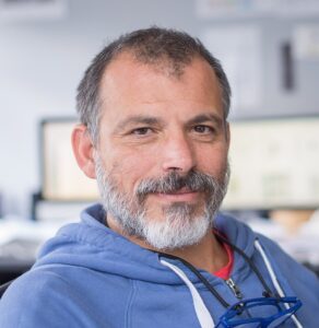
Gonzalo de Polavieja
Course Director
Champalimaud Foundation, PT
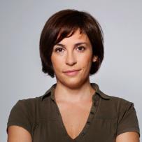
Kristin Branson
Course Director
HHMI Janelia, USA
Il Memming Park
Course Director
Champalimaud Foundation, PT
Keynote Speakers
Ann Kennedy – Northwestern University, US
N. Alex Cayco Gajic – École Normale Supérieure, France
Ben Cowley – Cold Spring Harbor, US
Kim Stachenfeld – DeepMind, UK
Jennifer Sun – Caltech, US
Josue Nassar – Stony Brook University, US
Laura Driscoll – Stanford University, US
Maneesh Sahani – Gatsby Computational Neuroscience Unit, UK
Nakul Verma – Columbia University, US
Patrick Mineault – xcorr, Canada
Rui Ponte Costa – University of Oxford, UK
Sara Solla – Northwestern University, US
Surya Ganguli – Stanford University, US
Instructors
Alice Robie – HHMI Janelia, USA
Brad Hulse – HHMI Janelia, USA
Joao Barbosa – Ecole Normale Superieure, France
Tiago Marques – Champalimaud Foundation, Portugal
André Mendonça – Champalimaud Foundation, Portugal
Course content
Programme
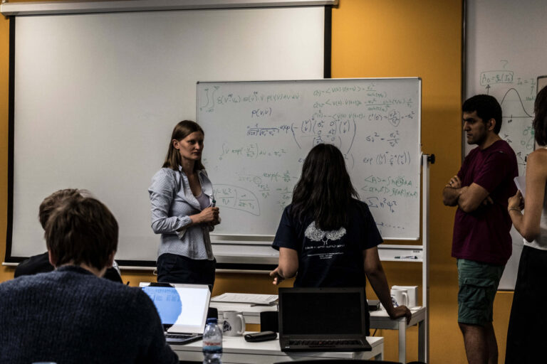
During the first two weeks, there will be lectures in the morning, and evening tutorials will consist of analytical and computational application of the concepts learned in the morning. The third week consists of lectures in the mornings and projects in the evening.
First week
The first week will consist of a mini-course in Machine Learning, especially those techniques used in Neuroscience. This will include supervised and unsupervised learning, learning for time series and Reinforcement Learning, as well as a day dedicated to practical aspects in the use of Machine Learning Techniques.
Day 1: History of AI and the relationship between Machine Learning and Neuroscience
Day 2: Supervised Learning
Day 3: Unsupervised Learning
Day 4: Practical consideration of the use of Machine Learning
Day 5: Learning for Time Series
Day 6: Reinforcement Learning
Second Week
The second week discusses how Machine Learning techniques are applied in Neuroscience. This will include the Machine Learning tools used to extract information from datasets of animal behavior and brain activity, Machine Learning models from brain activity and behavior, models using spiking neural networks and which are the limits ofML techniques.
Day 7: ML tools in animal behavior
Day 8: ML tools in brain activity
Day 9: Models derived from the brain and behavior
Day 10: ML-based models of the brain & discrepancies
Day 11: Theory-based modeling, and its connection to ML
Day 12: Bio-inspired ML/spiking networks
Third week
The third week has lectures on more advanced topics and evenings on projects.
Students will design projects applying machine learning (ML) methods to neural recording and animal behavior data sets. Neural recording data sets provided will be from a variety of techniques, including electrophysiology, calcium imaging, and fMRI. Behavior data sets provided will be from video. Students may focus on developing new ML algorithms for improving automated tracking, segmentation, categorization, or representation learning. Alternatively, they may focus on applying these ML methods to large data sets to discover new biological insights. A third option is to explore ML-based computational models of neural activity or behavior. Through these projects, students will gain hands-on experience using modern ML approaches, coding in PyTorch, and gain a deeper understanding of the power and potential failures of ML.
Datasets
Behavioral Datasets
- Fish Fish in groups, raw and tracked
- Some rodents and ants, raw and tracked
- Flies data from Maabe and other datasets.
Neural Datasets (spike trains)
- Neural Latent Benchmark ones
- Neuromatch ones (e.g. Steinmetz)
- Brain machine interface related datasets
- IBL datasets
Other Neural Datasets ( LFP, fMRI, etc)
Computational models (not datasets but used as input for further analysis or modeling)
- RNNs trained on tasks
- Models trained on fish in collectives
Techniques
-
Deep learning tools for analyzing complex behavior data
-
Neural manifolds
-
Modeling neural computation with recurrent neural networks
-
State space modeling of neural time series
-
Probabilistic modeling of neural data and behavior
-
Deep learning models of brain activity and behavior
-
Spiking neural networks
-
Best practices for high quality neuroscience research using machine learning
Champalimaud Centre for the Unknown, Portugal
The Champalimaud Foundation is a private, non-profit organization, established in 2005 and dedicated to research excellence in biomedical science. Completed in 2010, the Champalimaud Centre for the Unknown is a state-of-the-art centre that houses the Champalimaud Clinical Centre and the Champalimaud Research, with its three parallel programs – the Champalimaud Neuroscience Programme, the Physiology and Cancer Programme, and the Experimental Clinical Research Programme.
Initially focused on a system and circuit approach to brain function and behavior, the Centre expanded to incorporate molecular and cell biological expertise. The Centre comprises 26 research groups (circa 400 researchers) leading independent curiosity-based research.
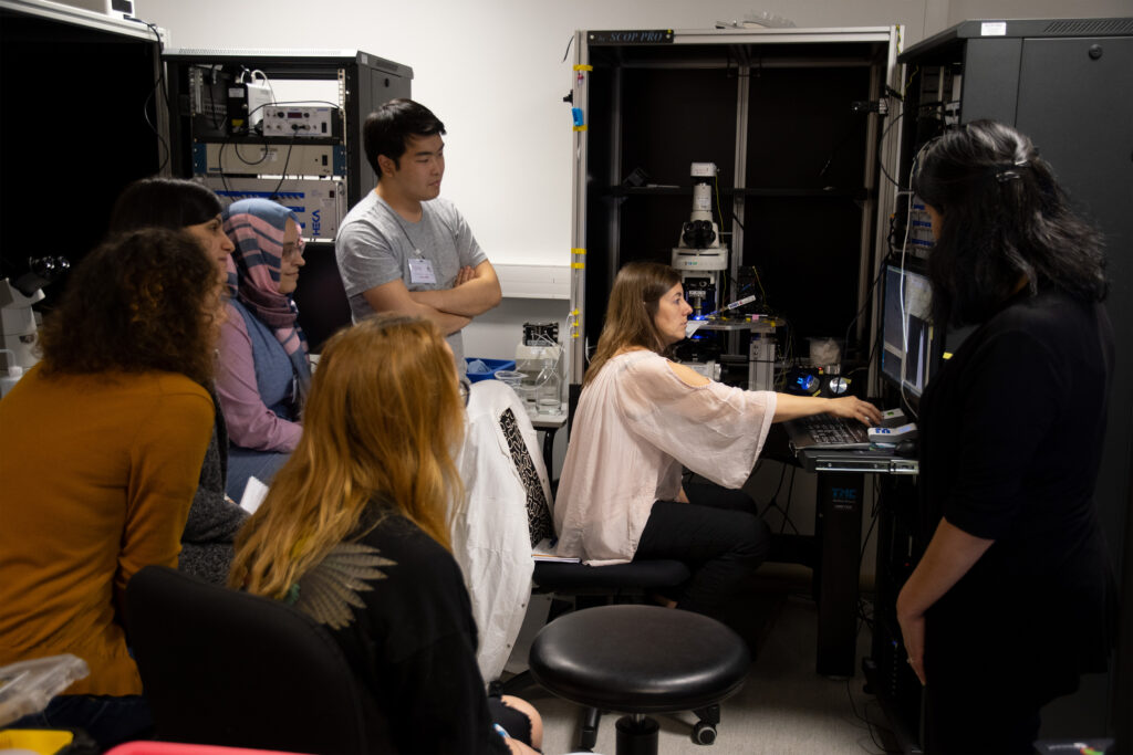
Facilities
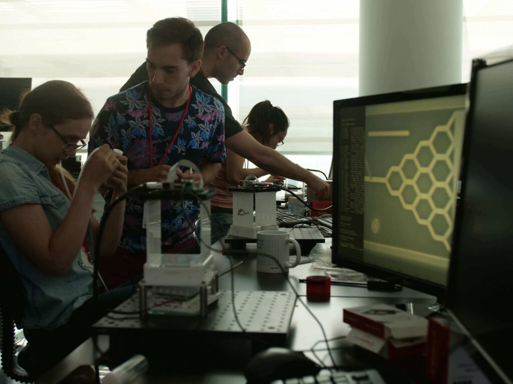
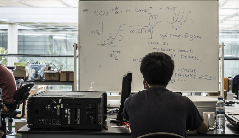
The Centre provides Facilities dedicated for Training, some in their entirety, for use by the CAJAL Advanced Neuroscience Training Programme. These include the Teaching Laboratory, a fully equipped open lab space for 20-30 students that can be dynamically reconfigured to support a full range of neuroscience courses. It also overlooks, via floor to ceiling windows, a tropical garden and the river. The experimental spaces include: Imaging Lab: A dark-room containing a full size optical table is used for advanced imaging setups (two-photon microscopy, SPIM, etc.) and custom (course-designed) optical systems.
Registration
Fee : €2.950,00 (includes tuition fee, accommodation and meals)
Applications closed on 23rd January 2023
The CAJAL programme offers 4 stipends per course (waived registration fee, not including travel expenses). Please apply through the course online application form. In order to identify candidates in real need of a stipend, any grant applicant is encouraged to first request funds from their lab, institution or government.
Kindly note that if you benefited from a Cajal stipend in the past, you are no longer eligible to receive this kind of funding. However other types of funding (such as partial travel grants from sponsors) might be made available after the participants selection pro- cess, depending on the course.
Sponsors