Course overview
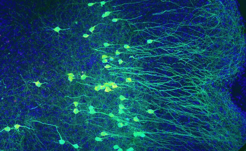
The course provides advanced training into the field of neural organoids for the modeling of brain development and physiopathology. Neural organoids, three-dimensional models derived from human pluripotent stem cells, have been revolutionizing the study of the human brain and its disorders by recapitulating key steps of human brain development including neural stem cells patterning, expansion, differentiation and connectivity. Moving beyond a basic introduction, this intensive course will focus on emerging frontiers research directions, equipping the next generation of neuroscientists with cutting-edge frontier know-how.
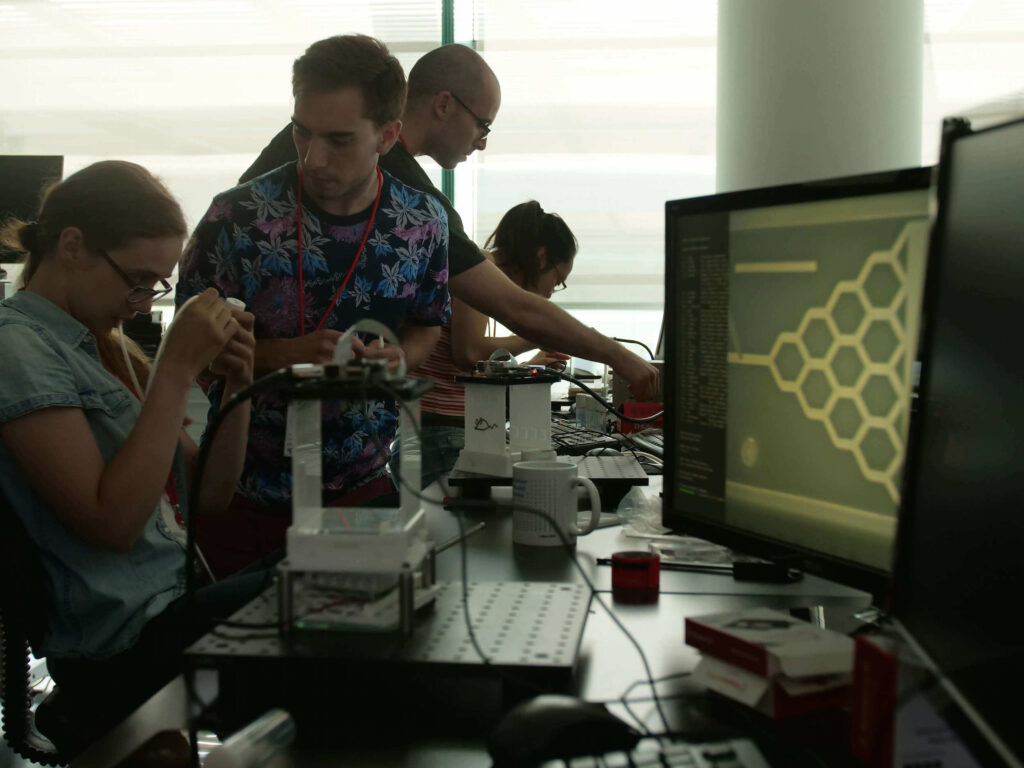
The course emphasizes an integrative, hands-on approach that bridges the gap between theoretical knowledge and practical applications by combining experimental and computational methods. Participants will gain a sophisticated theoretical framework and the practical insights to pursue the frontiers of neural organoid modelling in their own research.
Course Directors
Giuseppe Testa
University of Milan and Human Technopole, Italy
Flora Vaccarino
Yale University, USA
Scientific Directors
Nicolò Caporale
University of Milan and Human Technopole, Italy
Emanuele Villa
Human Technopole, Italy
Keynote Speakers
Juergen Knoblich, Institute of Molecular Biotechnology, Austria
Michael Wells, University of California, USA
Helena Kilpinen, University of Helsinki, Finland
Orly Reiner, Weizmann Institutre, Israel
Giulio Franzese, EURECOM, France
Pietro Michiardi, EURECOM, France
Jason Stein, University of North Carolina at Chapel Hill, USA
Carlo Colantuoni, Johns Hopkins University School of Medicine, USA
Alexej Abyzov, Mayo Clinic, USA
Sara Bizzotto, Institut Imagine, France
Instructors
Laila Sathe, University of California, USA
Puigdevall Costa, University of Helsinki, Finland
Jeyoon Bok, University of Michigan, USA
Giulio Franzese, EURECOM, France
Carlo Colantuoni, Johns Hopkins University School of Medicine, USA
Ariana Berenice Marquez Gonzalez, University of North Carolina at Chapel Hill, USA
Wang, Yifan, Mayo Clinic, USA
Natalia Baumann, Institut Imagine, France
Alexandre Jourdon, Yale School of Medicine, USA
Soraya Scuderi, Yale School of Medicine, USA
Davide Castaldi, Human Technopole, Italy
Manuel Lessi, Human Technopole, Italy
Luciana Isaja, Human Technopole, Italy
Filippo Mirabella, Human Technopole, Italy
Course content
Topics
This course will focus on five themes:
● Neural organoids for population-scale neurodevelopmental modelling to connect molecular and clinical phenotypes;
● Mosaic models to understand the molecular basis of clonal dynamics and non-cellautonomous effects in the human brain;
● Mathematical modelling of neural organoid biology;
● Morphogen patterning as a driver of brain region and cell lineage specification and a source of biases in brain morphogenesis;
● Somatic mutations in pluripotent lines to trace developmental lineages.
Aims and Techniques
The primary objective of this intensive course is to equip young neuroscientists with the sophisticated theoretical framework and practical expertise required to successfully implement neural organoids in their own research endeavors.
Upon completion of the course, participants will be able to:
● Demonstrate a comprehensive understanding of the principles of differentiation of
pluripotent stem cells into different neural organoid types.
● Critically assess the advantages and disadvantages of neural organoid modelling and select the most appropriate designs for their research questions.
● Master essential techniques for the characterization of neural organoids, including multiplexing approaches to scale-up modelling, integrating molecular and clinical endophenotypic scales, and dissecting unexplored dynamics of developmental systems.
● Design and execute experiments leveraging neural organoids to model complex physiopathological processes, through a combination of experimental and computational approaches.
● Establish a professional network with leading scientists and peers, fostering future collaborations and career development.
Bordeaux School of Neuroscience, France
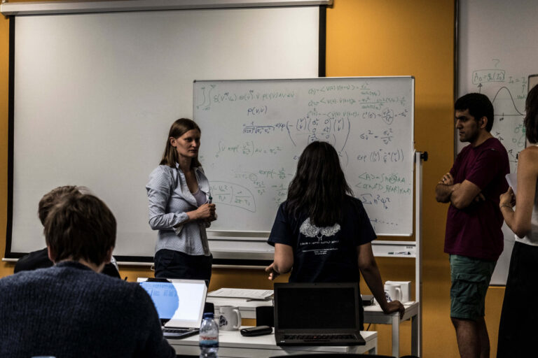
The Bordeaux School of Neuroscience is part of Bordeaux Neurocampus, the Neuroscience Department of the University of Bordeaux. Created in 2015, it is currently directed by David Perrais. Throughout the year, renowned scientists, promising young researchers and many students from any geographical horizon come to the School.
The school works on this principle: training in neuroscience research through experimental practice, within the framework of a real research laboratory.
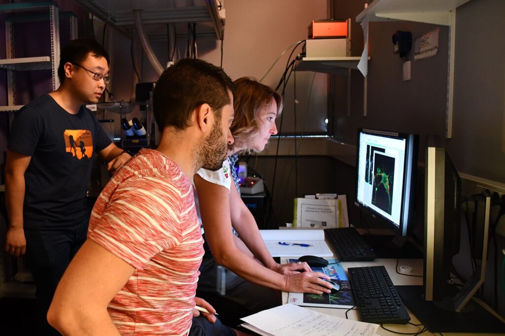
Facilities
Their dedicated laboratory (500m2), available for about 20 trainees, is equipped with a wet lab, an in vitro and in vivo electrophysiology room, IT facilities, a standard cellular imaging room, an animal facility equipped for behavior studies and surgery and catering/meeting spaces. They also have access to high-level core facilities within the University of Bordeaux. They offer their services to international training teams who wish to organize courses in all fields of neuroscience thanks to a dedicated staff for the full logistics (travels, accommodation, on-site catering, social events) and administration and 2 scientific managers in support of the experimentation.
Registration
Fee : 4.500 € (includes tuition fee, accommodation and meals)
Applications are now closed!
Sponsors